The injury
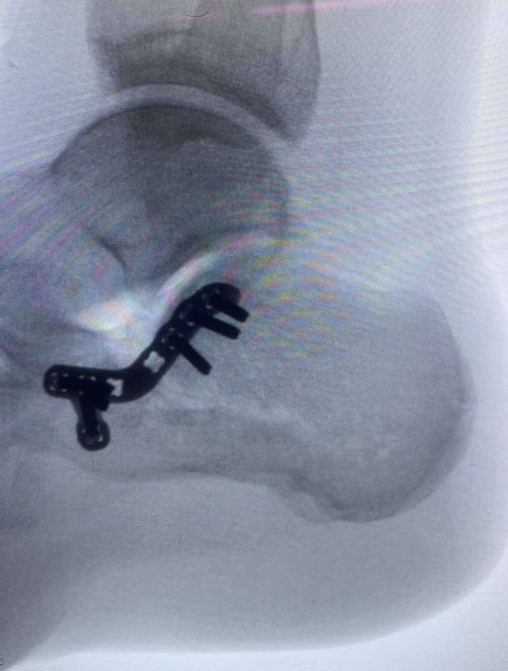
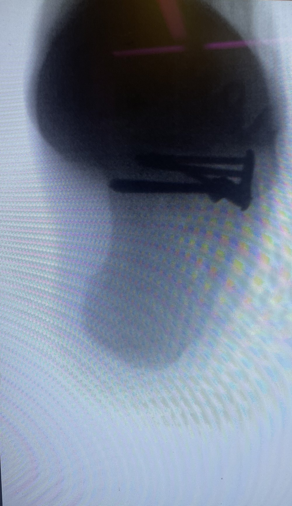
The calcaneus is typically fractured by axial load, such as a fall from height. The subtalar joint is usually involved and the heel becomes broadened and shortened.
Surgical approach
The traditional extensile lateral incision carries a high rate of wound complications. Where the fracture pattern allows, I use a sinus tarsi approach, reduce the joint under image guidance and hold it with a low-profile plate and screws.
Indications for surgery
Displaced intra-articular fractures in patients whose skin condition, vascular status and smoking history permit surgery to be undertaken safely. Undisplaced fractures are treated in a boot.
Recovery: points specific to this injury
Elevation for two weeks, six to eight weeks non-weight-bearing, then progressive loading. Swelling may persist for several months, and a proportion of patients subsequently require subtalar fusion for post-traumatic arthritis.
Recovery after surgery
The timeline below is a typical pathway after fracture surgery. The exact plan depends on the injury and the fixation, and will be confirmed at each review.
| Stage | What to expect |
|---|---|
| Weeks 0–2 | A temporary cast is applied and the leg must be kept non-weight-bearing. During this period the limb should be elevated, with the toes above the level of the nose, ideally for 55 minutes out of every hour, to help control swelling. You can mobilise with crutches, a knee scooter or an iWALK hands-free crutch (see recovery equipment). |
| At 2 weeks | If possible, the cast is changed to a boot so that soft-tissue management can begin: desensitisation, and moisturising of the scars. Creams such as Kelo-cote or castor oil can be used for wound care (see recovery equipment). If a boot is not yet appropriate, a further cast is applied from week 2 to week 6. |
| At 6 weeks | The cast is removed and you move into a boot, typically a short surgical boot where possible, or a Vacoped boot, depending on the type of surgery. |
| Physiotherapy | Physiotherapy starts as soon as the limb is accessible, which is either at 2 weeks or at 6 weeks. |
| Exercise | Core and upper-body work can start as soon as the cast has been removed and you are in a boot, which can be as early as week 2, but with nothing through the injured limb. From 6 weeks, gentle range-of-motion exercise can begin, such as a cross-trainer, a fixed (static) bike and swimming. Impact sports such as running and jumping can normally start at 12 weeks; this depends to some extent on the fracture configuration. |
| Longer term | These are often complex and severe injuries. Improvement can take up to 18 months to reach a new baseline. Swelling often persists for months, typically around 9 to 12 months, and some swelling often persists lifelong, without complete resolution of the ankle swelling. |
Risks of surgery
The risks associated with this surgery include, but are not limited to:
- Pain
- Bleeding
- Infection
- Scarring
- Damage to nerves and blood vessels
- Deep vein thrombosis (DVT) — a blood clot in the veins of the leg
- Pulmonary embolism (PE) — a blood clot that travels to the lungs, which can be life-threatening
- Stiffness
- Non-union — failure of the bone to heal
- Malunion — the bone healing in an incorrect position
- The need for further surgery, such as removal of metalwork
These risks are discussed individually before surgery.
First two weeks after surgery
Items patients often find useful in the first two weeks. Always follow your surgical team's instructions; these are general suggestions, not medical advice. As an Amazon Associate I earn from qualifying purchases.
Recovery and after your operation: helpful products
As an Amazon Associate I earn from qualifying purchases. These are general suggestions, not a substitute for advice from your surgical team.
Arranging an appointment. Private patients are seen at King Edward VII's Hospital, The London Clinic, Cromwell Hospital and HCA Healthcare at The Shard.
Call 020 7046 8004 or send an enquiry.
Radiographs are from my own practice, reproduced uncropped with identifying details removed. This page provides general information and is not a substitute for individual assessment.