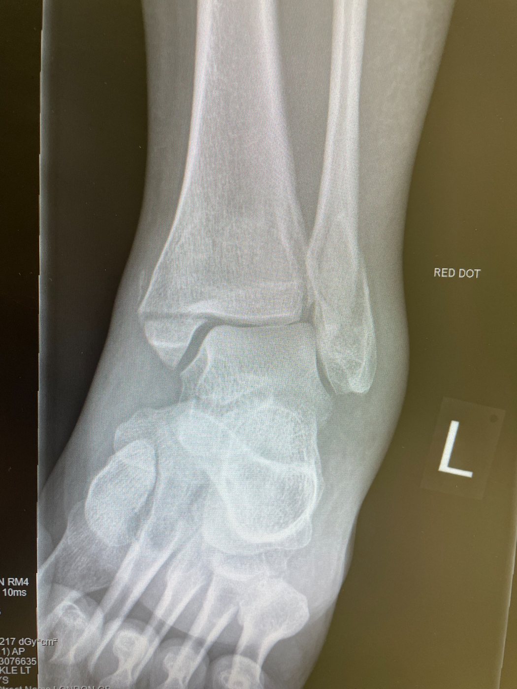
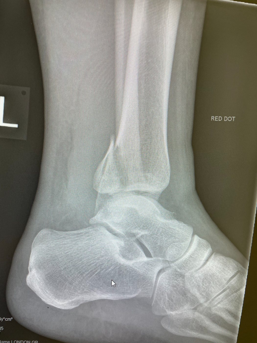
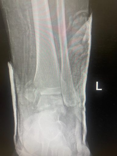
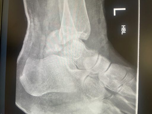
The injury
A fracture of the fibula, of the tibia at the medial or posterior malleolus, or of both. The ankle is a constrained hinge and functions well only when the joint surface is accurately reduced; displacement of one or two millimetres alters the way load is transmitted across the cartilage.
When surgery is advised
Stable fractures with the talus correctly positioned are treated in a boot and weight-bearing is begun early. Operative fixation is advised for unstable patterns: displacement of the talus, disruption of the syndesmosis, or a significant posterior malleolar fragment.
The operation
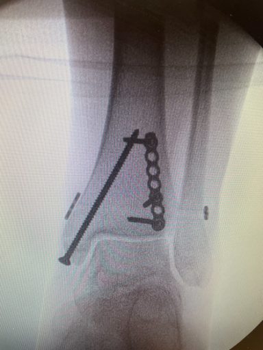
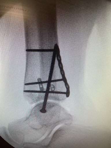
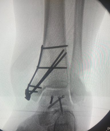
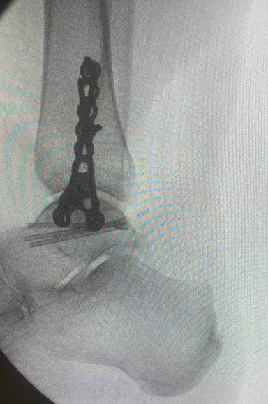
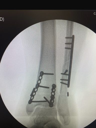
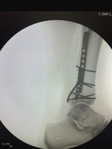
The fibula is reduced through a lateral incision and held with a contoured plate and screws. A disrupted syndesmosis is stabilised with screws or a suture-button device, and the medial and posterior malleoli with screws or a small plate. Most patients are discharged the same day or the following morning.
Recovery
Two weeks in a splint to allow the wound to heal, followed by a removable boot. The point at which weight-bearing is permitted depends on the fracture pattern and bone quality. Physiotherapy usually begins at around six weeks, and driving is generally possible at eight to ten weeks after a right ankle fracture. Metalwork is retained unless it becomes symptomatic.
Arranging an appointment. Private patients are seen at King Edward VII's Hospital, The London Clinic, Cromwell Hospital and HCA Healthcare at The Shard. NHS patients are seen at King's College Hospital.
Call 020 7046 8004 or send an enquiry.
Radiographs are from my own practice, reproduced uncropped with identifying details removed. This page provides general information and is not a substitute for individual assessment.